Wednesday 23 May 2012

Conference call


As I headed for some note taking I was stopped in my tracks by a new breed of students......

Wednesday 16 May 2012

Hidden beauties #15

Card 15 - Male shield fern


Ferns are one of my favourite plants.


Fern flora on a garden wall


Fascination of ferns

Tuesday 15 May 2012

Hidden beauties #14

Card 14 - Expulsion by teeth


Monday 14 May 2012

Hidden beauties #13

Card 13 - Catching by hairs


I seem to be meeting my self set blogging challenge to post every day (apart from weekends - a "rule" I set up en route). The cigarette cards have certainly provided food for thought, not least a realisation that there are projects I have had in my mind for a long time and have done nothing about. Seeing this picture of grass reminds me that I want to set up a series of pictures of  flowers we don't notice - grass flowers, tree flowers, things like plantain. Then there is Spirogyra and other pond life that are so easy to obtain and look at.

Tomorrow I am expecting the arrival of a new microscope, so no excuses ..... At the moment things are quite fraught with student support note taking, but that is only to last another week or so and then I can begin!

Friday 11 May 2012

Hidden beauties #12

Card 12 - Wonderful flower dust


Thursday 10 May 2012

Hidden beauties #11

Card 11 - A leaf slice


As can be seen from the following images, leaf structure can be quite varied, but the overall "plan" is quite similar. The upper epidermis protects the leaf and then underneath that is usually the palisade layer; this is the layer where most of the photosynthesis takes place, its cells are usually tall and cylindrical and well supplied with chloroplasts.. Beneath this layer are the more loosely packed and irregularly shaped cells of the spongy mesophyll layer; beneath that is the lower epidermis. Most of the stomata are usually found in this layer. Carbon dioxide enters the leaf through the stomata and diffuses through to the palisade cells to be combined with water (brought in from the root via the xylem tissue) to form carbohydrates using light energy from the sun.
Pampas grass - transverse section through leaf

Pinguilica (Butterwort) TS through leaf showing mucilage secreting hair which helps trap insects

Marram grass TS through leaf. Marram grass needs to conserve water so the leaf is curled, stomata are situated in pits,.

TS through the leaf of a xerophyte (a plant that lives in dry conditions) - its epidermis is thick to reduce water loss  by transpiration, its stomata are in pits which are surrounded by hairs to increase humidity around them and so reduce water loss by transpiration.

More information can be found in this article I wrote for Microscopy UK

Wednesday 9 May 2012

Hidden beauties #10

 Card 10 - An airing apparatus


Hmmm, rather a broad sweeping statement to say that pondweed has no external beauty!

Potamogeton transverse section through stem

The image above is another one I took from a prepared microscope slide, again, the colouration is due to stains used to make tissues more obvious. The air spaces which keep the plant buoyant are clearly visible. Buoyancy is necessary to keep the leaves near the surface so that photosynthesis can be maximised. The dark structure in the middle is the vascular tissue, this central arrangement gives the stem more flexibility in moving water. In non woody land plants the vascular tissue is often arranged near the edge of the stem to form a strengthening cylinder; in contrast, root vascular tissue is arranged centrally, thus providing more strength against pulling forces.

Tuesday 8 May 2012

Hidden beauties #9

Card 9 - Pith stars


This image of stellate parenchyma is from one of many prepared slides given to me. The slides had been made as part of a university course and provided a basis for my first explorations into microscopy. The slides themselves seemed ancient, having been made before I was born.

The photograph is one of my first forays into photomicrography. As a biology teacher I had realised that pupils very often were not sure what they were looking for on a slide - for example, air bubbles were often mistaken for cells.  So, I decided that if I could provide photographs which helped them identify what they were looking for, the learning experience might be more fulfilling.

The set up with my original microscope was quite challenging, the lens from the SLR was removed and then using a special attachment, the camera was set on the eyepiece. Focusing was quite difficult, both because the light entering the camera wasn't too bright (it was reflected from an old table light via a mirror) and also because the weight of the camera made the whole set up rather unsteady. 

Monday 7 May 2012

Hidden beauties #5, #6, #7 and 8

Circulation within cells,  and plant hairs


Card 5 -  A pink and hairy skin


Card 6 - Circulation currents in hairs


Card 7 - Rotation


Card 8 - A miniature forest


The cytoplasmic streaming is called cyclosis, I've found this video which demonstrates it:


Plant hair variety.


Viewing under polarised light can make the hairs more obvious

Nettle sting

Hairs on a seed, I haven't been able to remember the name of the seeds, but I do know they came with warnings!


Friday 4 May 2012

Plant movement

The cucumber plant we have just bought provided inspiration yesterday.


A bit of time lapse photography.

Hidden beauties #4

Card 4 -The "mouths" of leaves


The stomata are the pores through which gas exchange takes place.The two guard cells surrounding each stoma contain cholorophyll (unlike the other cells in the epidermis); it is the guard cells that control the size of the stomata.

A "cast" can be taken from the surface of leaves using nail varnish. When the nail varnish is peeled off it can be put under the microscope.
Nail varnish cast of leaf epidermis from a dicotyledon (top) and monocotyledon (bottom)
More images can be see in an article I wrote for 1997 in Microscopy UK (http://www.microscopy-uk.org.uk/mag/indexmag.html?http://www.microscopy-uk.org.uk/mag/art97b/epider.html) (This article now looks very dated, html and web page design having moved on incredibly in the intervening time!)


The following images are of prepared epidermal tissue on a microscope slide, the blue colour is due to the dye used to make structures stand out more clearly.

Prepared slide: leaf epidermis, dicotyledon (top), monocotyledon (bottom)
Commelina leaves are ideal for observing stomata as their epidermis peels very easily. The following image is one I took from a leaf of a plant I grew from seed. The chloroplasts are clearly visible in the guard cells.


Thursday 3 May 2012

Hidden beauties #3


Card 3 - "Green stars" and "half bricks"


The "sugar makers" are chloroplasts, the cell organelles containing chlorophyll which  carry out photosynthesis (making food from carbon dioxide and water, using light energy).

A bit of word derivation: Zygnema is from the Greek, zygon - yoke and nema - thread.

It's been a long time since I did any pond dipping, maybe a future project would be to collect some pond scum, put it under the microscope and shine lights from different directions and to see if the chloroplast movement is observable.


Something completely different... I am reading Diane Ackerman's The moon by whale light, in which she says that it is the temperature of the eggs that determines the sex of baby crocodiles. This I knew, but what I hadn't considered is that as sex is not determined genetically, crocodiles don't have sex chromosomes.

Wednesday 2 May 2012

Hidden beauties #2

Card 2 - A plant with spiral bands


Spirogyra, there are almost 400 species of this genus. I can visualise the slide I had of this alga, to the naked eye just a tangle of pale green threads, but under the microscope the spiral arrangement of chloroplasts was very clear.

Click here to see some images

Tuesday 1 May 2012

Hidden beauties #1


I have set myself a blogging challenge ...

In our upheaval, various things have come to light, amongst them some sets of cigarette cards I bought from ebay several years ago. My plan had been to do something with them, that plan can now come to fruition.

My challenge is to photograph each card and then post some extra material.


Cigarette cards apparently go back to the 1890s when card was inserted in the then paper packets to protect the contents. Manufacturers realised the potential the card had as an advertising ploy, then, to maintain product loyalty, sets of pictorial sequences began to appear.


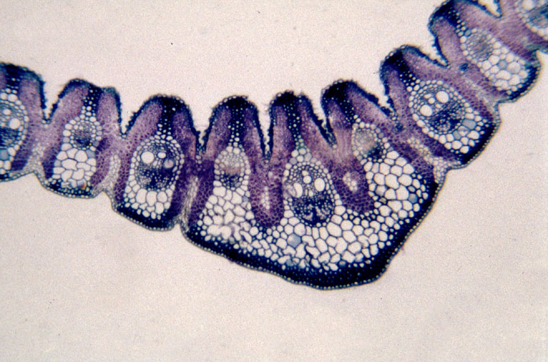
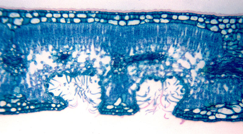
Card 1 - Hidden Beauties


I got my first microscope when I was sixteen soon after I decided that biology was the subject I really wanted to study. It was a monocular with no internal light source, light was transmitted by reflection though the mirror at its base.

I remember some of the first things I looked at were bees in various degrees of dissection. To actually see the hook and groove arrangement that enables their wings to beat in synchrony was amazing